nanoSilver helps to increase the survival rate for white shrimp when infected with WSSV
Silver nanoparticles enhance vannamei survival against white spot syndrome virus (WSSV) by activating the shrimp immune system.
DESCRIPTION
The global aquaculture industry has been steadily growing over the past decade. However, the aquaculture industry also faced many difficulties, causing damage to the whole industry. One of the problems facing the industry is the epidemic, typically white spot syndrome virus (WSSV). Many research efforts have been made, one of which is a study carried out by a number of scientists, studying the ability of nanosilver particles to promote immune response. shrimp disease infected with WSSV. The results of the scientists showed that, with a low dose of silver nanoparticles of about 12ppb (0.012ppm), the survival rate of infected shrimp increased by 20%.
The scientists found that an increase in the survival rate of infected shrimp by 20% when treated with silver nanoparticles was associated with a 2-fold increase in LGBP gene expression. LGBP (Lipopolysaccharide and beta-1,3-glucan binding protein) is a key gene in shrimp immune system when infected with WSSV virus causing white spot disease in shrimp.
The excess iron in alum-contaminated ponds facilitates the growth of the white spot virus. The results showed that a small dose of silver nanoparticles was able to enhance the immune response in shrimp to fight white spot disease, the use of silver nanoparticles had no adverse effects on healthy shrimp. The disease is caused by the virus, so there is currently no special treatment, which is completely based on the immune system of the infected shrimp and improving the living environment. Therefore, to prevent white spot disease caused by WSSV virus, it is advisable to combine the use of nanosilver and control so that iron (iron alum) is not present in the culture medium.
Experimental description of nano silver
The studied shrimp sample is a batch of shrimp with a size of about 8g (125 shrimp / kg). Farmed in tanks at about 26 degrees Celsius. Shrimp are maintained under these conditions until they reach 10 g (100 shrimp / kg). Shrimp were fed with a conventional commercial feed with 35% protein.
Inoculate the WSSV white spot disease virus provided by ITVY Aquaculture Laboratory (Mexico) into the 3rd abdomen of shrimp.
Silver nanoparticles (AgNP) were tested at the concentrations of 120ppm and 12ppm under different conditions as in Table 2 (Table 2) below.
– Group S1 was tested with nanosilver concentration of 120ppm on shrimp samples infected with white spot virus.
– Group S2 was tested with nano silver concentration of 12ppm on shrimp samples infected with white spot virus.
– Group S3 was tested with nanosilver concentration of 120ppm on shrimp samples infected with white spot virus, in iron containing medium (FeSO4).
– Group S4 was tested with the concentration of nano silver is 12ppm on shrimp samples infected with white spot virus, in iron containing medium (FeSO4).
– S5 group was tested on shrimp samples infected with white spot virus but without nano silver and iron (FeSO4).
– Group S6 was tested on normal healthy shrimp samples, without using nano silver and in iron-free environment (FeSO4).
4. Analysis of virus causing WSSV white spot disease and gene expression of the immune system by real-time PCR method. The Vp24 gene of the WSSV white spot virus encodes the main protein for viral expression located on the envelope of the virus.
Nano silver test results
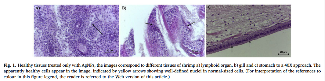
The results of tissue analysis of shrimp Photographs of healthy tissues exposed to nano silver were tested on the placenta tissues of shrimp: a) lymphoid organ, b) gill and c) stomach as shown in figure 1 below. Photographs showed that nanosilver used at the same concentration as research did not cause any adverse effects.
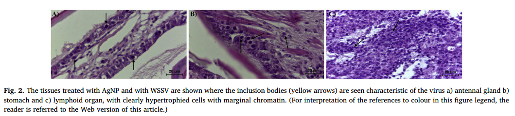
On the other hand, shrimp infected with white spot virus WSSV (group S5) showed an enlarged cell nucleus or an enlarged organ. The results are similar to the iron samples of groups S3 and S4 with the appearance of nano-silver in figure 2 below. Imaging results were not significantly different when shrimp were treated with nano-silver and not treated. What is special, however, is that life time is different when treated with nanosilver.
2. Tested with WSSV virus
The test samples (S1-S5) were again exposed to the white spot virus. Then the death rate was recorded after 6 hpi (hours post-infection: the number of hours after viral infection) until the S5 sample was recorded (without using nano silver) completely dead. The results are shown in figure 3 below:
The results in Figure 3 show that the 100% mortality rate with the S5 sample not treated with nano silver, after 96 hours of viral infection Groups S3 and S4 are two test groups with the presence of iron is the black and green line with the highest mortality rate in comparison to the remaining test samples. It proves that the environment in the presence of iron creates favorable conditions for the virus, causing the mortality rate to increase rapidly. The final results showed that the group S5, S3 and S4 had the mortality rate of 100% after 96 hours of infection. However, the S1 and S2 groups are the two groups using nanosilver, the mortality rate drops to 10% and 20%, ie the death rate of group S1 is 80% and the S2 group is 90%. The S6 control group was a healthy group with a 0% mortality rate. Completely identical test conditions and no cross-contamination determined by realtime PCR method.
3. Results of viral content on test samples
Determination of the content of virus causing white spot disease in shrimp through protein of Vp24 gene of WSSV virus, determined through realtime PCR method. The results are shown in Figure 4C below.
Results showed that the groups S1 – S4 had significantly more virus than the S5 group. Although the S1 – S4 groups are the nano silver treated groups. The S5 group is the nanosilver untreated group.
4. Results of shrimp immune system response to white spot virus
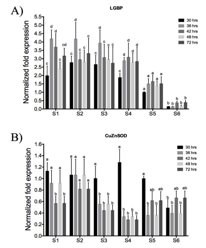
The response of shrimp immune system to white spot virus infection is determined through protein of LGBP and CuZnSOD gene segments. The results are shown in Figures 4A and 4B as shown below.
The results in Figure 4A show that, with samples S1 and S2, the activity of the LGBP gene segment reached its maximum level at 36 hours of WSSV infection. After 36 hours, LGBP gene activity decreased to normal levels. For samples S3 and S4, similar results are compared to samples S1 and S2 even in the environment of samples S3 and S4, there is the presence of iron. Thus, samples from S1 – S4 using nano silver have a positive effect on the immune boost through stimulation of the LGBP gene.
In contrast to the immunological enhancement of the samples from S1 – S4, the S5 sample has a very low immune response, only half of that of the samples from S1 – S4. The reason is that S5 sample was infected with WSSV white spot virus but did not use nano silver.
Sample S6 is a sample that is not infected with WSSV white spot virus and does not use nano silver, the body’s immune response is very low because there is no pathogen. Show that nanosilver particles support immune response, not the direct cause of LGBP gene activation.
The results in Figure 4B show that, in black column after 30 hours from the time of infection, the samples from S1 – S5 group have similar values and twice that of sample S6 (reference sample is not infected). It was shown that nano silver used for samples from S1 – S4 did not affect Cu, Zn-SOD genes. This is due to the fact that the S6 model is not infected. After 36 hours, the gene’s activity returned to normal levels, only for samples S3 and S4, due to the presence of iron in the samples S3 and S4. That showed that, when shrimp infected, CuZn-SOD gene was active in the first 30 hours, after 36 hours, it was normal. In an iron-containing medium, it reduces the activity of this gene, increases the mortality rate of shrimp.
REFERENCES
Alba R. Ochoa-Meza, Ana R. Álvarez-Sánchez, Carlos R. Romo-Quiñonez, Aarón Barraza, Francisco J. Magallón-Barajas, Alexis Chávez-Sánchez, Juan Carlos García-Ramos, Yanis Toledano-Magaña, Nina Bogdanchikova,Alexey Pestryakov, Claudio Humberto Mejía-Ruiz, Silver nanoparticles enhance survival of white spot syndrome virus infected Penaeus vannamei shrimps by activation of its immunological system, Fish and Shellfish Immunology 84 (2019) 1083–1089.